An electromyogram (EMG) is a common clinical test used to assess function of muscles and the nerves that control them. EMG studies are used to help in the diagnosis and management of disorders such as the muscular dystrophies and neuropathies. Nerve conduction studies that measure how well and how fast the nerves conduct impulses are often performed in conjunction with EMG studies.
Examples of EMG studies (courtesy of Seward Rutkove, MD, Department of Neurology, Beth Israel Deaconess Medical Center/Harvard Medical School) are given here.
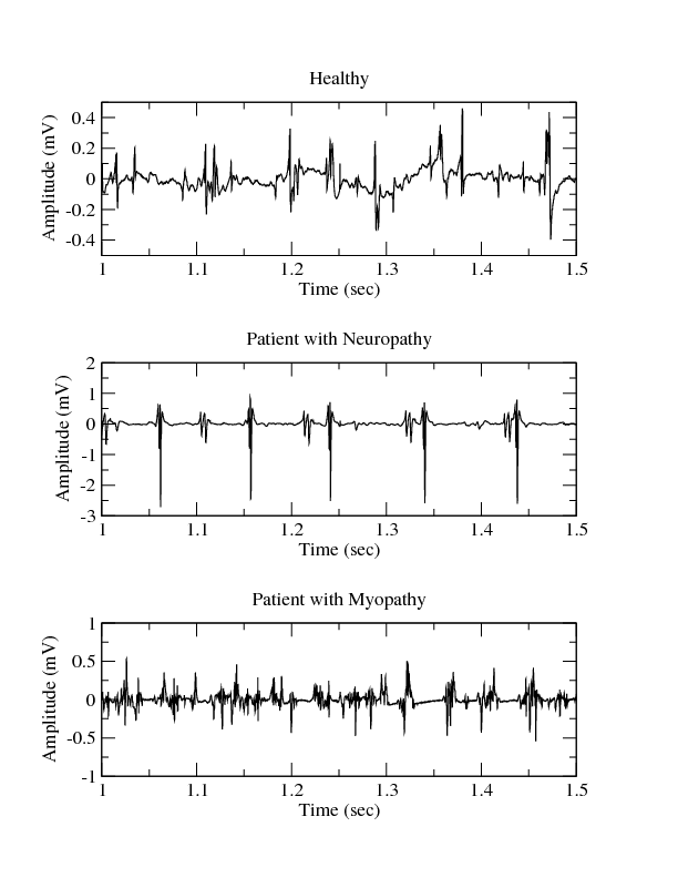
Data were collected with a Medelec Synergy N2 EMG Monitoring System (Oxford Instruments Medical, Old Woking, United Kingdom). A 25mm concentric needle electrode was placed into the tibialis anterior muscle of each subject. The patient was then asked to dorsiflex the foot gently against resistance. The needle electrode was repositioned until motor unit potentials with a rapid rise time were identified. Data were then collected for several seconds, at which point the patient was asked to relax and the needle removed.
The figure shows three examples of EMG data from: 1) a 44 year old man without history of neuromuscular disease; 2) a 62 year old man with chronic low back pain and neuropathy due to a right L5 radiculopathy; and 3) a 57 year old man with myopathy due to longstanding history of polymyositis, treated effectively with steroids and low-dose methotrexate. The data were recorded at 50 KHz and then downsampled to 4 KHz. During the recording process two analog filters were used: a 20 Hz high-pass filter and a 5K Hz low-pass filter.
Suggested readings about EMG:
- Kimura J. Electrodiagnosis in Diseases of Nerve and Muscle: Principles and Practice, 3rd Edition. New York, Oxford University Press, 2001.
- Reaz MBI, Hussain MS and Mohd-Yasin F. Techniques of EMG signal analysis: detection, processing, classification and applications. Biol. Proced. Online 2006; 8(1): 11-35.
Name Last modified Size Description
Parent Directory -
emg_neuropathy.txt 26-Apr-2010 19:58 2.1M
emg_neuropathy.dat 26-Apr-2010 20:20 289K digitized signal(s)
emg.png 05-Sep-2009 18:46 41K
RECORDS 26-Apr-2010 21:15 40 list of record names
DOI 21-Sep-2015 19:00 19
|
If you would like help understanding, using, or downloading content, please see our Frequently Asked Questions. If you have any comments, feedback, or particular questions regarding this page, please send them to the webmaster. Comments and issues can also be raised on PhysioNet's GitHub page. Updated Friday, 28-Oct-2016 22:58:42 CEST |
PhysioNet is supported by the National Institute of General Medical Sciences (NIGMS) and the National Institute of Biomedical Imaging and Bioengineering (NIBIB) under NIH grant number 2R01GM104987-09.
|